Rev. Ciencias Veterinarias, Vol. 39, N° 1, [1-8], E-ISSN: 2215-4507, enero-junio, 2021
DOI: https://doi.org/10.15359/rcv.39-1.1
URL: http://www.revistas.una.ac.cr/index.php/veterinaria/index
Short Communication: First report of the trematode Platynosomum illiciens (Trematoda: Digenea) in Felis catus in Costa Rica, Central America
Comunicación corta: Primer reporte del trematodo Platynosomum illiciens en Felis catus en Costa Rica, América Central
Comunicação curta: Primeiro relato do trematódeo Platynosomum illiciens em Felis catus na Costa Rica, America Central
Brandon Castillo Azofeifa1, Valeria Zamora Segura1, Francinie Quirós Padilla1, Emilia Vindas van der Wielen1, Francisco Valerio Solís1, Takashi Iwaki2, Sugiyama Hiromu3, Ana Jiménez Rocha4 , Juan Alberto Morales-Acuña1
 Corresponding author: Ana Jiménez, Email: ana.jimenez.rocha@una.ac.cr
1 Servicio de Patología, Escuela de Medicina Veterinaria, Universidad Nacional, Heredia, Costa Rica. E mail: bcastillo1101@gmail.com, valzamora16@gmail.com, chiniqp@hotmail.com, evindas_10@hotmail.com, franvalerios96@hotmail.com, juan.morales.acuna@una.ac.cr
2 Meguro Parasitological Museum, Tokyo, Japan. Email: iwaki@kiseichu.org
3 National Institute of Infection Diseases, Department of Parasitology, Tokyo, Japan.
Email: hsugi@nih.go.jp
4 Laboratorio de Parasitología, Escuela de Medicina Veterinaria, Universidad Nacional, Heredia, Costa Rica.
Email: ana.jimenez.rocha@una.ac.cr
Received: May 5, 2020 Corrected: January 27, 2021 Accepted: February 2, 2021
Abstract
This paper is the first description of the trematode P. illiciens found in a domestic feline (Felis catus) in Costa Rica, detected as an incidental finding at necropsy. The cat’s carcass was accepted for a necropsy in the Pathology Service of the Veterinary Medicine School of the National University, Costa Rica. However, the origin and clinical history of this cat are unknown. Adult P. illiciens flukes were detected in the feline’s gallbladder. Species identification was conducted by morphological characterization and molecular sequencing analysis. Assessing the possible foci of this trematode in the country could be a useful tool to understand its cycle in Costa Rican mammals and birds and consider it as an organism capable of infecting and affecting the domestic feline population in the country.
Keywords: Central America, trematode, Digenea, feline.
Resumen
Este artículo es la primera descripción del trematodo P. illiciens encontrado en un felino doméstico (Felis catus) en Costa Rica, detectado como hallazgo incidental en la necropsia. El cadáver del gato fue aceptado para una necropsia en el Servicio de Patología de la Escuela de Medicina Veterinaria de la Universidad Nacional. Sin embargo, se desconoce el origen y la historia clínica de este gato. Se detectaron gusanos adultos de P. illiciens en la vesícula biliar del felino. La identificación de la especie se realizó mediante caracterización morfológica y análisis de secuenciación molecular. La evaluación de los posibles focos de este trematodo en el país podría ser una herramienta útil para conocer su ciclo en los mamíferos y aves costarricenses y considerarlo como un organismo capaz de infectar y afectar a la población de felinos domésticos del país.
Palabras clave: América Central, trematodo, Digenea, Felino.
Resumo
Este artigo é a primeira descrição do trematódeo P. illiciens encontrado em um felino doméstico (Felis catus) na Costa Rica, detectado como um achado incidental em necropsia. O cadáver do gato foi aceito para necropsia no Serviço de Patologia da Escola de Medicina Veterinária da Universidade Nacional. No entanto, a origem e a história médica deste gato são desconhecidas. Vermes adultos de P. illiciens foram detectados na vesícula biliar felina. A identificação da espécie foi realizada por meio de caracterização morfológica e análise do sequenciamento molecular. A avaliação dos possíveis focos deste trematódeo no país pode ser uma ferramenta útil para entender seu ciclo em mamíferos e aves da Costa Rica e considerá-lo como um organismo capaz de infectar e afetar a população felina doméstica do país.
Palavras-chave: América Central, trematódeo, Digenea, Feline
Introduction
The species of trematodes belonging to the genus Platynosomum are parasites that affect both mammals and birds. The niche inside these hosts includes the biliary system, liver, and pancreas. This genus has a worldwide distribution and a complex taxonomy because of its great morphological variation (Pinto et al., 2014; Pinto et al., 2016). Three scientific species names have been used throughout time to describe these trematodes: P. concinnum, P. fastosum, and P. illiciens (Pinto et al., 2018). Previous studies by Rodriguez (1963), quoted in Pinto et al. (2018), found no significant differences in morphology between P. illiciens and P. fastosum, although the former was detected from birds and the latter from mammals. However, Pinto et al. (2018) mentioned that it is incorrect to use the name P. concinnum, originally described as Dicrocoelium concinnum; the first scientific name used for the trematode found in cats because of the morphological description of D. concinnum does not adjust to Platynosomum diagnosis. The study conducted by Pinto et al. (2018) gives an interesting finding to resolve the taxonomic issue about P. illiciens and P. fastosum, because the ITS1 sequences (KU987672–KU987674) showed high similarity between them, confirming that P. illiciens is conspecific with P. fastosum. According to Pinto et al. (2016), both species are unrelated to cryptic species, and the members of the family Dicrocoeliidae, which includes the genus Platynosomum, have a low host specificity, which could explain why P. illiciens is found in both birds and mammals.
Platynosomiasis is a common disease in tropical and subtropical areas (Lenis et al., 2009). In Latin America, P. illiciens is the most common liver fluke in domestic cats; the parasite invades and parasitizes the gallbladder, intrahepatic and extrahepatic bile ducts, liver, and sometimes the small intestine, pancreatic ducts, and lungs. Patients with chronic infections can present various clinical signs, including jaundice, vomiting, diarrhea, weight loss, depression, and even death. However, these symptoms are nonspecific, which often makes rapid diagnosis and treatment difficult (Lenis et al., 2009). The life cycle is indirect and involves two intermediate hosts.
The first host is terrestrial snails that ingest the operculated eggs containing miracidium; the second host is a terrestrial isopod that eats the sporocysts containing cercariae develop in the snail or ingested by several species of amphibians or lizards where they encyst as metacercariae in the bile duct or gallbladder. A cat ingests these hosts, and the metacercariae will excyst and migrate to the cat’s bile duct where they mature. Lizards or amphibians could act as second intermediate hosts and as paratenic hosts, and they will have the metacercariae encysted in the gallbladder and bile duct (Bowman, 2014). Birds, felines, mustelids, canids, marsupials, and primates have been registered as definitive hosts, and experimentally rodents have acted as definitive hosts as well, getting the infection by eating amphibians or reptiles containing the metacercariae (Bowman, 2014; Lenis et al., 2009).
In Costa Rica, P. illiciens has been reported in the areas of Puerto Viejo de Sarapiquí, Heredia, and Tilarán, Guanacaste (Brenes & Arroyo, 1962). Its presence has been determined only in birds; the only species reported (five cases) is the Leucopternis semiplumbeus, also known as the semiplumbeous hawk. The semiplumbeous hawk is a bird of prey that lives permanently in Costa Rica. Its diet consists of other birds, small lizards, and rodents. They inhabit the Caribbean side of the country, including Tortuguero and Puerto Viejo de Sarapiquí in Heredia province (Henderson, 2010). P. illiciens has been found in the gallbladder of the semiplumbeous hawk (Brenes & Arroyo, 1962).
The morphological description and sequence analysis of adult P. illiciens detected from a cat liver is given. This report constitutes the first confirmation of P. illiciens isolated from animals other than birds in Costa Rica.
Materials and methods
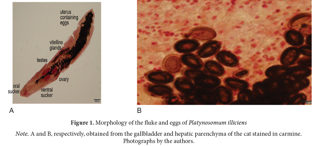
The corpse of a feline was accepted for a necropsy by the Pathology Service of the Veterinary Medicine School of the Faculty of Health Sciences, at the National University of Costa Rica. The cat was a male mongrel with short white and yellow hair and unknown age (approximately 8 years old). His body weight was 3.4 kg but showed a low body condition score with unknown clinical history and origin. At necropsy, the cat presented multifocal pulmonary congestion and hemoptysis, as well as hydrothorax and hydropericardium. When examining the abdominal cavity, severe chronic diffuse fibrinous active peritonitis due to the jejunum’s rupture and leakage of intestinal contents into the abdominal cavity was found. As an incidental finding, small parasites were found in the gallbladder and hepatic parenchyma (in the intrahepatic bile duct), collected on a slide and then analyzed morphologically and molecularly. The detected trematodes were fixed in 70% ethanol, stained with borax carmine, and mounted in Canada balsam. The morphology of one specimen was observed in detail using a microscope (Figure 1). The identification at the generic level was made using identification keys by Pojmańska (2008). The molecular analysis of the adult fluke was performed by DNA isolation, amplification of the nuclear ribosomal ITS2 region of about 470 base pairs (bp) by PCR, and sequencing of the amplicon by the methods described before (Sugiyama et al., 2013). The primers used for PCR amplification and sequencing were as follows:

3S (forward, 5ʹ-GGTACCGGTGGATCACTCGGCTCGTG-3ʹ (Bowles et al., 1995), and A28 (reverse, 5ʹ-GGGATCCTGGTTAGTTTCTTTTCCTCCGC-3ʹ (Blair et al., 1997).
The PCR amplification was performed with 0.25 µM of respective primers and 2.5 U of Phusion High-Fidelity DNA Polymerase (New England Biolabs, Ipswich, Massachusetts, USA). The PCR amplification was performed on a thermal cycler (TaKaRa PCR Thermal Cycler Dice Gradient, Takara Bio Inc., Shiga, Japan), with 30 cycles of 98°C for 10 sec, 55°C for 10 sec, and 72°C for 15 sec. An initial denaturation and final extension were carried out at 98°C for 30 sec and at 72°C for 7 min, respectively. The amplification product was sequenced either with the forward or reverse primer and the BigDye Terminator v3.1 Cycle Sequencing Kit (Thermo Fisher Scientific, Waltham, Massachusetts, USA) on an automated sequencer (3730xl DNA Analyzer, Thermo Fisher Scientific). Sequences were aligned and compared using GENETYX-Win software (Ver. 13, Genetyx Co., Tokyo, Japan).
Results
Three adult flukes and eggs from the gallbladder and hepatic parenchyma of the cat were detected, with no findings indicating a significant lesion in the liver.
The measurements of the adult flukes were as follows (in micrometers, unless otherwise stated):
- Body slender and elongate, whole length 7.56 mm; maximum width 1.05 mm.
- The size of the oral and ventral sucker is 450 × 414 and 487 × 451, respectively.
- Two large and elliptic testes (737 × 303 and 640 × 309) are positioned posterior to the ventral sucker in parallel.
- The ovary is elliptic (498 × 262), positioning posterior to one of the testes, submedian.
- Vitelline bands are arranged in lateral fields, expanding from the level of the ovary to the 3/5 of body-length.
- The uterus expands the hindbody and contains numerous brown oval eggs (30–35 × 21–23).
- The genital pore is positioned at the level of intestinal bifurcation.
According to previous reports, the morphology of the trematode corresponds with those of P. illiciens (Nguyan et al., 2017; Pinto et al., 2016).
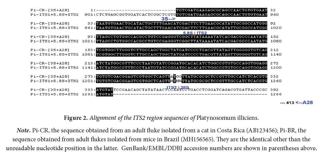
The amplicon’s sequence analysis revealed that the generated product was 413 bp (without primer sequences). The sequence obtained was identical to that of P. illiciens (DDBJ/EMBL/GenBank accession number: MH156565) occurring from Brazil (Pinto et al., 2018), as shown in Figure 2. Hence, the nucleotide sequence obtained in this study has been deposited as the adult P. illiciens prevalent in Costa Rica in the DDBJ/EMBL/GenBank database under accession number of LC500530.

The necropsy findings of the cat did not reflect significative lesions in the liver, but the presence of the parasite P. illiciens was confirmed by the molecular identification (PCR) of the eggs and adult worms collected in the bladder and bile duct.
Discussion
This paper is the first description of the trematode P. illiciens found in a domestic cat in Costa Rica. The morphology and molecular analysis were carried out, but a limitation of the study is not having obtained the amplicon; then, rewriting the morphological description is a recommendation for future studies.
During the necropsy, it was determined that a possible cause of death of the feline was severe chronic peritonitis caused by a rupture of the jejunum. Adult P. illiciens flukes were detected from the gallbladder and hepatic parenchyma, probably from the intrahepatic bile duct, and trematode eggs were detected from the hepatic parenchyma. However, although no significant macroscopic changes were detected in the liver, and it was not possible to confirm these findings at a histopathological level, the possibility that the trematode caused microscopic alterations, altering thus the hepatic and biliary function, cannot be discarded. In this cat, it is possible that the number of trematodes parasitizing the liver was not enough for the feline to show specific symptoms and macroscopic findings.
Bowman (2014) stated that cats acquire the infection with P. illiciens by eating lizards, toads, and geckos infected with the metacercariae. However, Pinto et al. (2014) experimentally demonstrated that lizards are not obligatory second intermediate hosts for P. illiciens but can function as paratenic hosts. Isopods harboring metacercariae can play a role for direct transmission to the definitive hosts. The cat from the present case might become infected with P. illiciens by eating either the paratenic host such as lizards, toads, and geckos, or the second intermediate host such as isopods.
The provenance of the feline, where P. illiciens were found, is unknown, but the only published report of this trematode in the country was in 1962, in birds from the Costa Rica’s Caribbean coast and Guanacaste (Brenes & Arroyo, 1962). This raises the question if subdiagnosis of the infection is taking place in the country, considering the fact that the cat was brought from a site of the Greater Metropolitan Area, so more cases of domestic felines with nonspecific clinical signs that could be infected by platynosomiasis may be confused with other conditions, and therefore, are not being treated properly. The presence of P. illiciens is unknown in other countries of Central America; it is very important to do research on this.
In Latin America, P. illiciens has been found in domestic cats in several countries. Colombia is among the countries that have reported P. illiciens in cats (Felis catus). At first, trematodes found in a stray cat from Turbo, Antioquia, were identified as Dicrocoelium dendriticum; however, further investigation and taxonomic analysis proved that the trematodes found actually were P. illiciens (Lenis et al., 2009). Similarly, P. illiciens has been reported in domestic cats in Brazil. In this country, the prevalence of Platynosomum spp. varies depending on the state; in São Paulo, the incidence reported ranges from 1.07% to 5.6%, whereas in Rio de Janeiro, the prevalence reached up to 56.25% (Salomão et al., 2005). In another study conducted in Brazil, the percentage of infection of P. illiciens in domestic cats found in the city of Araguaína, Tocantins, was 33.3% (Gomes-de Oliveira Sobral et al., 2019).
Further studies are necessary to clarify the distribution range of this trematode in Costa Rica; the information that can be collected could be a useful tool to understand the life cycle of P. illiciens in mammals and birds. The first and second intermediate hosts and paratenic hosts have not been investigated in this country either. Additional work is needed to answer these epidemiological questions, which could identify the infection sources of P. illiciens to wild and domestic animals and, then, establish preventive measures against platynosomiasis. The presence of P. illiciens is unknown in other countries of Central America; it is very important to do research on this.
Considering that this tends to be an asymptomatic disease or that cats develop nonspecific clinical signs, veterinarians are urged to consider platynosomiasis as a differential diagnosis in cases of suspected liver and biliary disease in felines; thus, they can better approach and treat the condition.
Conclusion
The presence of the parasite Platynosomum illiciens in a domestic feline in Costa Rica is of great relevance for the veterinary practice, as it allows to suggest more studies to determine if the finding was accidental or if there is a national prevalence in the domestic feline population. Learning about the life cycle of this parasite and its presence in intermediate, paratenic, and definitive hosts, as well as its prevalence in the country, is very important to understand and prevent the pathologies that may occur as a result of an infection.
Acknowledgment
We thank Dr. César Salas Pérez for his support and the National Institute of Infection Diseases, Department of Parasitology in Tokyo for providing the stained histopathological specimens of the cat. This study was partially supported by JSPS KAKENHI Grants (25305011 and 16H05820).
Conflict of interests
The authors declare that they have no conflicts of interest.
References
Blair, D., Agatsuma, T., Watanobe, T., Okamoto, M. & Ito, A. (1997). Geographical genetic structure within the human lung fluke, Paragonimus westermani, detected from DNA sequences. Parasitology, 115(4), 411-417. doi: 10.1017/S0031182097001534
Bowles, J., Blair, D. & McManus, D.P. (1995). A molecular phylogeny of the human schistosomes. Molecular Phylogenetics and Evolution., 4(2), 103-109. doi: 10.1006/mpev.1995.1011
Bowman, D.D. (2014). Georgi’s Parasitology for Veterinarians (10th Edition). Elsevier, Missouri.
Brenes, R.R. & Arroyo, G. (1962). Helmintos de la República de Costa Rica XX: Algunos tremátodos de aves silvestres. Revista de Biología Tropical, 10(2), 205-227.
Gomes de Oliveira, M.C., Pereira, S.A., Peixoto, T.M., Rocha, S., Martins, R., da Silva, R.A., Silva, T., Ferreira, F.E. & Dias, H. (2019). Infection by Platynosomum illiciens (= P. fastosum) in domestic cats of Araguína, Tocantins, northern Brazil. Revista Brasileira de Parasitologia Veterinária., 28(4), 786-789. doi: 10.1590/s1984-29612019070
Henderson, C.L. (2010). Birds of Costa Rica: A field guide. University of Texas Press, United States.
Lenis, C., Navarro, J.F. & Velez, I. (2009). First case of platinosomosis from Colombia: Platynosomum illiciens (Digenea: Dicrocoeliidae) in Felis catus, Turbo, Antioquia. Revista Colombiana de Ciencias Pecuarias, 22(4), 659-66.
Nguyen H.M., Van Hoang H. & Ho, L.T. (2017). Platynosomum fastosum (Trematoda: Dicrocoeliidae) from cats in Vietnam: morphological redescription and molecular phylogenetics. Korean Journal of Parasitology, 55(1), 39–45. doi: 10.3347/kjp.2017.55.1.39
Pinto, H.A., Mati, V.L. & Melo, A.L. (2014). New insights into the life cycle of Platynosomum (Trematoda: Dicrocoeliidae). Parasitology Research, 113(7), 2701-2707. doi: 10.1007/s00436-014-3926-5
Pinto, H.A, Mati, V.L. & Melo, A.L. (2016). Can the same species of Platynosomum (Trematoda: Dicrocoeliidae) infect both mammalian and avian hosts? Journal of Helminthology, 90(3), 372–376. doi: 10.1017/S0022149X15000152
Pinto, H.A., Pullido-Murillo, E.A., Braga, R.R., Mati, V.L.T., Melo, A. L. & Tkach, V.V. (2018). DNA sequences confirm low specificity to definitive host and wide distribution of the cat pathogen Platynosomum illiciens (= P. fastosum) (Trematoda: Dicrocoeliidae). Parasitology Research, 117(6), 1975-1978. doi: 10.1007/s00436-018-5866-y
Pojmańska, T. (2008). Family Dicrocoeliidae Looss, 1899. In: Bray, R.A., Gibson, D.I. & Jones, A. (Eds.). Keys to the Trematoda (Volume 3, pp. 233–260). CAB International and Natural History Museum, London.
Salomão, M., Souza-Dantas, L.M., Mendes-de-Almeida, F., Branco, A.S., Bastos, O.P.M., Sterman, F. & Labarthe, N. (2005). Ultrasonography in hepatobiliary Evaluation of domestic cats (Felis catus, L., 1758) infected by Platynosomum Looss, 1907. The International Journal of Applied Research in Veterinary Medicine, 3(3), 271-279.
Sugiyama, H., Singh, T.S. & Rangsiruji, A. (2013). Paragonimus. In: Liu, D.Y. (Ed.). Detection of Human Parasitic Pathogens (pp. 423-436). CRC Press, Florida, USA.

Artículo publicado con Licencia Creative Commons, Atribución-No-Comercial, SinDerivadas 3.0 Costa Rica